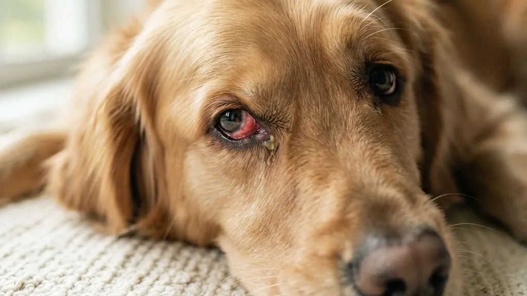
Dog eye infection is the phrase most owners reach for the moment they see redness or goop, yet the label is wrong more often than it is right. A red, weepy, squinting eye can be bacterial, but it can just as easily be an allergy, dry eye, a trapped grass seed, or acute glaucoma that will destroy vision in hours. The American Kennel Club and the Merck Veterinary Manual both stress the same thing: the tissue involved and the type of discharge matter more than the word you use. This guide separates the true infection from its look-alikes, gives you a timed urgency triage, and hands you the actual test values a veterinarian uses.
Quick answer: A dog eye infection usually shows thick yellow-green discharge, redness, and mild squinting, and it responds to a diagnosed antibiotic ointment. But the same eye can hide an emergency. A bulging eye, a cornea that turns cloudy or blue suddenly, a fixed dilated pupil, or intense pain points toward acute glaucoma or a corneal ulcer, not a simple infection. When those signs appear, skip home care and get to a vet the same day. Confirming the cause needs a Schirmer tear test, fluorescein stain, and eye-pressure reading before any medication.
What a real dog eye infection is, and its five common impostors
A dog eye infection means a pathogen, usually a bacterium such as Staphylococcus or Streptococcus, has colonized eye tissue and triggered inflammation with thick, colored discharge. That is the narrow, accurate definition. The problem is that most owners apply the word to any unhappy eye, and the majority of unhappy eyes are something else. Getting the category right is the whole game, because the wrong home remedy can blind a dog.
The Merck Veterinary Manual groups eye problems by the tissue affected, and each tissue tells a different story about cause and danger. Conjunctivitis affects the pink membrane lining the eyelid and eyeball. Blepharitis is the eyelid skin itself, often crusty and swollen. Keratitis involves the clear cornea, the layer that can scar or ulcerate. Uveitis reaches internal structures like the iris, and it often signals a systemic disease elsewhere in the body. Dacryocystitis is an infection of the tear ducts, and it shows up as pus that seems to well up from the inner corner of the eye rather than the surface.
The five impostors that most often get mislabeled:
- Allergy: both eyes, watery clear discharge, itch and rubbing, rarely painful. It waxes with pollen and dust rather than turning yellow.
- Dry eye (keratoconjunctivitis sicca, KCS): thick, ropey, sticky discharge from too little tear film, often mistaken for pus. It is a tear-production failure, not a germ.
- Foreign body: usually one eye, sudden onset, hard squinting, tearing after a walk through tall grass.
- Glaucoma: a hard, painful, sometimes bulging eye with a cloudy cornea and a dilated pupil, driven by rising pressure.
- Corneal ulcer: intense pain, light sensitivity, and a surface defect that stains under fluorescein.
Viral causes exist too, including canine distemper and canine herpesvirus, along with fungi and parasites such as Demodex. Fungal eye disease is uncommon in dogs compared with cats, but it does turn up in dogs with weakened immune systems or after a penetrating injury that introduces spores. Demodex mites rarely cause eye trouble on their own, but they show up alongside facial skin disease in young dogs and can inflame the eyelid margins enough to look like blepharitis.
If your dog’s eye trouble comes with paw-chewing or skin flare-ups, the underlying driver may be allergic rather than infectious, and our guide to a dog paw yeast infection signs and treatment walks through how allergy and yeast overlap across the body. Dogs with generalized skin allergies frequently rub their faces on furniture and carpet, and that mechanical irritation alone can turn a mild allergic eye into a secondary bacterial one within a day or two, which is one reason the categories blur in real cases.
The differential table: match discharge, pain, and eye count to a cause
The single most searched question is not how to treat an infection. It is what this look means when the owner cannot yet name the problem. Owners cannot triage what they cannot categorize, so start with three observable facts: the type of discharge, whether the eye is painful, and whether one or both eyes are involved. Cross those against the cornea’s appearance and you narrow six possibilities to one or two fast.
This table condenses the patterns the Merck Veterinary Manual and VCA Animal Hospitals describe across their separate pathology pages into one view.
| Likely cause | Discharge | Pain / eyes | Cornea look |
|---|---|---|---|
| Bacterial infection | Yellow-green, thick | Mild, one or both | Usually clear |
| Allergy | Clear, watery | Itchy, both | Clear |
| Dry eye (KCS) | Thick, ropey, sticky | Dull ache, both | Dull, may be hazy |
| Foreign body | Watery then mucky | Sharp, one eye | May be scratched |
| Glaucoma / ulcer | Variable | Intense, often one | Cloudy or blue |
Read the table as a starting filter, not a diagnosis. Two clues override everything: pain and a cloudy cornea. A truly painful eye, or one whose cornea has gone hazy or bluish, jumps to the front of the queue regardless of discharge color. That combination is what separates a nuisance from an emergency, and it is the reason a phone photo is not enough. Even experienced owners misjudge cornea clarity in poor light.
Why discharge color is a clue, not a verdict
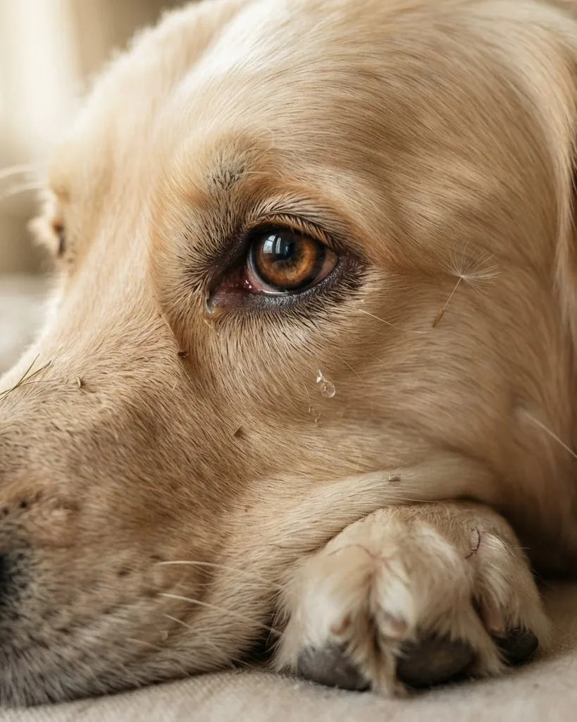
Yellow-green pus leans bacterial, and clear watery fluid leans allergic, but KCS produces thick sticky strands that mimic pus while carrying no infection at all. That single overlap is why so many infections are actually dry eye, and why an antibiotic drop alone fails to fix them. A dog can also have two problems layered on top of each other, for example a dry eye that has become secondarily infected because reduced tear film lets bacteria settle in and multiply. In that case the discharge color shifts from ropey and clear-ish to yellow over a few days, and treatment has to address both the underlying dryness and the new bacterial layer at once.
A worked example: reading your dog’s eye like a vet would
Picture a 7-year-old Cocker Spaniel with both eyes slightly red for a week, thick sticky discharge each morning, no obvious pain, and a dull look to the cornea rather than a bright shine. Using the table above, the pattern points toward dry eye rather than infection: bilateral, sticky rather than pus-like, low pain, hazy cornea. A Schirmer test confirms it, coming back at 7 mm per minute, well under the 10 mm per minute diagnostic cutoff for KCS. The plan becomes cyclosporine drops and artificial tears, not an antibiotic course, and the owner should expect this to be a long-term routine rather than a short fix.
Now compare that with a 3-year-old Labrador who comes in from the yard squinting hard in one eye only, pawing at the face, with clear watery tearing that turns slightly mucky within an hour. That pattern, one eye, sudden, sharp pain, matches a foreign body far more than an infection. Fluorescein stain often reveals a corneal scratch from the seed or debris that caused the reaction. The fix here is removal of the foreign material by a professional, pain control, and a short course of antibiotic ointment to prevent secondary infection while the scratch heals, not a wait-and-see approach.
A third case makes the urgency point sharply: an older Beagle whose eye suddenly looks bulged, with a cornea that has gone cloudy overnight and a pupil that will not react to light. Given the breed’s documented glaucoma predisposition, this profile should trigger a same-hour visit without waiting to see if it improves, because pressure readings in acute glaucoma commonly exceed 40 to 60 mmHg and vision loss accelerates with every hour of delay.
Urgency triage: within the hour, or watch at home
Sort the situation before you do anything else. Same-hour emergencies are those that threaten vision now: a bulging or proptosed eye, a cornea that turned cloudy or blue suddenly, a fixed dilated pupil, obvious intense pain, or apparent vision loss. Those signs point toward acute glaucoma or a perforating ulcer, and the Merck Veterinary Manual classifies acute glaucoma as a genuine ophthalmic emergency because pressure damage races ahead by the hour.
Use these tiers:
- Within the hour: bulging eye, sudden cloudy or blue cornea, fixed dilated pupil, severe pain, vision loss. Assume glaucoma or perforating ulcer until proven otherwise.
- Otherwise stable but unexamined: a case that looks uncomfortable but stable, without any of the same-hour warning signs above, and that has not been examined yet.
- Watch at home briefly: mild clear watering after a dusty walk, no pain, both eyes, dog acting normal. Recheck in a few hours.
The numbers behind the top tier are stark. Normal canine intraocular pressure sits around 15 to 25 mmHg, with some devices reporting a 10 to 25 mmHg range, according to Cornell University College of Veterinary Medicine and the Merck Veterinary Manual. In acute glaucoma the pressure typically climbs above 40 to 60 mmHg at presentation. Once pressure crosses 40 to 50 mmHg the eye needs emergency treatment because each hour raises the risk of permanent blindness. That is the difference between a drop from the pharmacy and a dash to the clinic.
A practical tip: photograph the eye in consistent daylight so you have something to compare against later. Comparing photos side by side often reveals whether swelling or discharge is stable, improving, or creeping worse, which is a better signal than memory alone. If the appearance visibly worsens between two photos taken half a day apart, move the case up to the same-hour tier rather than waiting out the original window.
If the eye trouble arrives alongside other worrying signs, treat the whole dog, not just the eye. Heavy nighttime breathing or restlessness can signal pain or systemic illness, and our explainer on what nighttime panting in dogs means covers how discomfort shows up after dark.
The tests a vet runs, and the values they are looking for
Diagnosis is not guesswork, and the values are specific enough that you can follow along at the appointment. Three bedside tests carry most of the weight: the Schirmer tear test measures tear production, fluorescein stain reveals corneal ulcers, and tonometry reads eye pressure. Each produces a number with a threshold, and those thresholds decide the treatment. This is where the top consumer articles fall short, because they describe symptoms without ever telling you what a normal reading is.
The Schirmer tear test is the anchor. Normal canine tear production runs above 15 mm per minute on the standard test, a threshold reported by the American College of Veterinary Ophthalmologists, Cornell University College of Veterinary Medicine, and Today’s Veterinary Practice. A value below 15 mm per minute is suggestive of KCS, and below 10 mm per minute is diagnostic of it. So an eye pouring out thick discharge with a Schirmer result of 6 mm per minute is not infected in the ordinary sense. It is starving for tears, and an antibiotic will not touch the cause.
A clinical study of 156 eyes across 78 dogs sharpened those lines. The normal group averaged a Schirmer result of 22.75 plus or minus 3.88 mm per minute, while the KCS group averaged 6.10 plus or minus 4.44 mm per minute. The same study measured tear film break-up time at 25.82 plus or minus 5.47 seconds in normal eyes versus 10.78 plus or minus 4.23 seconds in KCS eyes. Dry eye is not rare, either: in one study, 4 percent of dogs showed tear wetting below 15 mm per minute on the standard Schirmer test.
Then comes pressure. Tonometry gives the intraocular reading, and against the normal 15 to 25 mmHg band, a spike above 40 to 50 mmHg flips the visit from routine to emergency. Fluorescein stain settles the ulcer question by glowing green wherever the corneal surface is broken. Culture and sensitivity testing identifies the exact bacterium when a stubborn infection resists first-line drops. Together these four tests explain why a same-day exam beats any home remedy.
What the appointment actually looks like, step by step
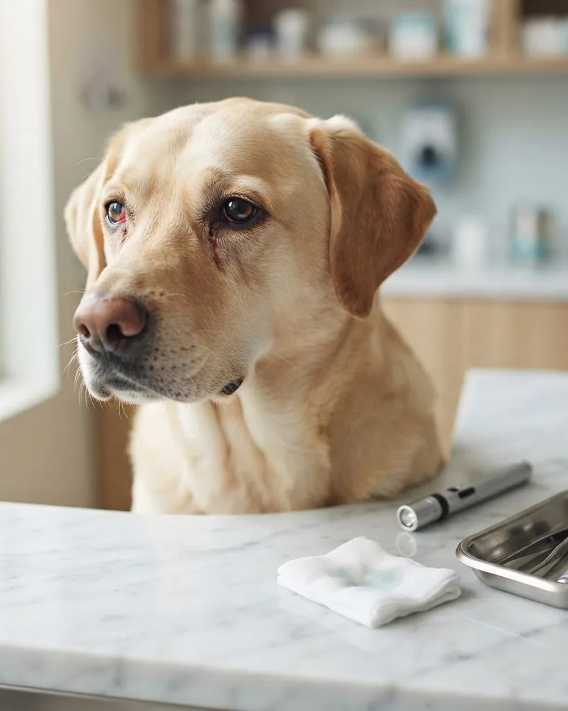
Most first ophthalmic exams follow a predictable order, and knowing it removes some of the anxiety of a same-day visit. The vet typically starts with a general look at both eyes side by side under bright light, comparing symmetry before touching either one. Next comes the Schirmer test, a small paper strip placed inside the lower eyelid for one minute to measure tear wicking, done before any drops are applied since anesthetic or dilating drops would skew the reading.
After the Schirmer strip is read, fluorescein stain goes in next, a drop of orange dye that glows green under blue light wherever the corneal surface is broken. Tonometry follows, using a handheld device pressed briefly against the numbed cornea to read pressure in mmHg. If the pattern still is not clear, a swab for bacterial culture may be taken before any treatment begins, since starting antibiotics first can make later culture results unreliable. The whole sequence usually takes under twenty minutes for a straightforward case.
Treatment by cause, and the home moves that make things worse
Treatment follows the diagnosis, never the symptom, because the same red eye demands opposite drugs depending on the cause. Bacterial infections get antibiotic drops or ointment. Viral causes, such as those linked to distemper or canine herpesvirus, are managed supportively or with antivirals rather than antibiotics. KCS is a lifelong tear-stimulation problem, and VCA Animal Hospitals lists topical cyclosporine or tacrolimus plus lubricants as the mainstay, frequently for the dog’s life. Structural problems like eyelid abnormalities or tumors need surgery, and systemic disease driving the eye must be treated at its root.
Blepharitis, the eyelid inflammation, leans on physical care. VCA Animal Hospitals recommends warm compresses held for 5 to 15 minutes several times a day, combined with gentle removal of crusted secretions. That simple routine does more for inflamed lids than any drop, and it costs nothing but time.
Glaucoma treatment is its own category because of the time pressure involved. Emergency pressure-lowering medication comes first, aimed at protecting remaining vision within hours rather than days. Depending on how the eye responds and whether vision is already lost, longer-term options range from ongoing pressure-control drops to surgical procedures that reduce fluid production inside the eye. Because primary glaucoma in predisposed breeds like Beagles often eventually affects the second eye too, a vet will usually recommend preventive monitoring or treatment for the unaffected eye once one eye is diagnosed.
Corneal ulcers split into simple and complicated categories. A simple, shallow ulcer often heals within days with topical antibiotic drops and an Elizabethan collar to stop rubbing. A deep or infected ulcer, especially one that is enlarging or has a melting appearance, can need surgical stabilization to prevent the eye from perforating. This is one of the clearest reasons why a corticosteroid should never go into an eye before an ulcer has been ruled out with fluorescein stain, since a steroid actively slows the collagen repair the cornea needs.
The home mistakes are the dangerous part. Do not do these:
- Never use a leftover human steroid eye drop on an undiagnosed eye. If a corneal ulcer is present, a corticosteroid can deepen it toward perforation.
- Never dig out a foreign body by hand. Manual removal can drive it deeper or scratch the cornea.
- Never bring hydrogen peroxide near the eye. It is not an eye rinse and it burns ocular tissue.
Gentle saline flushing and keeping the dog from rubbing are reasonable holding measures while you arrange a visit. An Elizabethan collar, even a soft fabric version, is worth putting on the moment you notice heavy rubbing or pawing, since a single hard scratch across an already inflamed cornea can turn a mild case into a surgical one. If the eye discharge coincides with a skin sore the dog keeps licking, the two problems may share an allergic root, and our walkthrough on the dog hot spot treatment and warning signs shows how quickly a moist lesion can escalate without care.
Breed predispositions and prevention that actually moves the needle
Some dogs are built for eye trouble, and knowing your breed’s weak spot changes how fast you react. Brachycephalic breeds, the flat-faced dogs with prominent eyes, expose more corneal surface to injury and drying simply because of their skull shape. Cocker Spaniels, Bulldogs, Cavalier King Charles Spaniels, Pugs, and Shih Tzus skew toward dry eye and cherry eye. Primary glaucoma clusters in Beagles, Cocker Spaniels, Basset Hounds, and Norwegian Elkhounds, which raises the stakes on any red, painful eye in those breeds.
The genetics are documented, not folklore. According to the Merck Veterinary Manual, Beagles and Norwegian Elkhounds develop primary open-angle glaucoma through a mutation in the ADAMTS10 gene. For an owner of one of those breeds, a sudden hard or cloudy eye is a same-hour event by default, because the odds of glaucoma are stacked before symptoms even start.
Prevention is unglamorous and effective. Keep the hair around the eyes trimmed so it cannot wick discharge or scratch the cornea. Wipe away debris with plain saline. Manage seasonal allergies before they escalate into constant rubbing. Control systemic conditions that spill into the eyes.
A few practical habits reduce risk further. Checking a brachycephalic dog’s eyes daily takes under a minute and catches early redness before it becomes painful. Trimming facial hair away from the eye margin every few weeks prevents hairs from scratching the cornea on windy walks. For breeds prone to KCS, a baseline Schirmer test done at a routine annual exam, even before symptoms appear, gives the vet a healthy number to compare against later if trouble develops, which speeds up diagnosis considerably.
Grooming choices matter here too, since harsh products around the face can inflame sensitive tissue, and our comparison of a hypoallergenic dog shampoo and its real limits explains what a gentle formula can and cannot prevent near the eyes.
Is a dog eye infection contagious to people?
Most canine eye conditions do not transmit to humans, so you can comfort a squinting dog without much worry for yourself. Some viral causes can pass between dogs, which matters in multi-dog homes. The American Society for the Prevention of Cruelty to Animals and general veterinary guidance both frame routine hand washing as sufficient household precaution.
What a visit costs you in time and follow-up
The real price of an eye problem is measured in follow-up, not just the first visit. A bacterial case caught early may resolve in one round of drops. KCS is the opposite. Because cyclosporine or tacrolimus therapy is usually lifelong, the commitment is measured in years, not days, and the payoff is preserving vision that would otherwise erode. That contrast is why the initial diagnosis is worth doing properly the first time rather than cycling through guesses.
Budget your attention around the tiers already described. An emergency-tier eye needs pressure control fast, since the window before glaucoma damage sets in is counted in hours once pressure passes 40 to 50 mmHg. A standard infection tolerates a next-day appointment. A mild watery eye can wait a few hours of observation. Matching your urgency to the tier keeps you from both panic and dangerous delay.
Follow-up visits also differ by cause in ways worth planning for. A simple bacterial conjunctivitis usually needs only a recheck near the end of the antibiotic course to confirm resolution. A corneal ulcer needs more frequent rechecks, often every few days at first, since fluorescein staining has to be repeated to confirm the defect is shrinking rather than growing. KCS needs periodic Schirmer retesting, typically at follow-up exams over the following months, to confirm the tear-stimulating medication is working and to adjust the dose if production remains low.
Common mistakes owners make when judging an eye problem
A few judgment errors come up again and again, and each one delays proper care. The first is assuming that because a dog is still eating and playing normally, the eye cannot be serious. Pain tolerance varies enormously between dogs, and a stoic dog with early glaucoma can still greet you at the door even as pressure climbs toward damaging levels.
The second common error is treating any yellow discharge as automatic proof of bacterial infection and starting a leftover antibiotic ointment without a vet visit. Old ointment can be expired, may not match the actual bacterium present, and delays a proper culture if the case does not respond. The third mistake is waiting to see if a cloudy cornea clears on its own overnight. Corneal cloudiness that appears suddenly rarely resolves without intervention, and the delay itself is the single biggest controllable risk factor for permanent vision loss in glaucoma cases.
Frequently asked questions
How do I know if my dog’s eye issue is an infection or dry eye?
Discharge alone cannot tell you, because dry eye produces thick sticky strands that look like pus. The deciding test is the Schirmer tear test. Normal tear production runs above 15 mm per minute, per the American College of Veterinary Ophthalmologists and Cornell University College of Veterinary Medicine. Below 10 mm per minute confirms dry eye, which needs tear-stimulating medication rather than antibiotics.
When is a dog’s red eye a true emergency?
Treat it as same-hour urgent if the eye bulges, the cornea turns cloudy or blue suddenly, the pupil is fixed and dilated, pain is intense, or vision seems lost. These suggest acute glaucoma or a perforating ulcer. In acute glaucoma, pressure often exceeds 40 to 60 mmHg at presentation, and each hour above 40 to 50 mmHg raises the risk of permanent blindness.
Can I use human eye drops on my dog?
No, not without a diagnosis. Human corticosteroid drops are especially dangerous, because on an undiagnosed eye with a corneal ulcer they can deepen the wound toward perforation. Plain saline rinsing is the only safe holding measure. Everything medicinal should follow a veterinary exam that includes fluorescein staining to rule out an ulcer first.
Which breeds get eye problems most often?
Flat-faced breeds like Pugs, Bulldogs, and Shih Tzus face higher exposure injury and dry eye. Cocker Spaniels and Cavalier King Charles Spaniels tend toward tear disorders. For glaucoma, Beagles, Basset Hounds, and Norwegian Elkhounds carry elevated risk, and the Merck Veterinary Manual ties Beagle and Norwegian Elkhound glaucoma to an ADAMTS10 gene mutation.
Is a dog eye infection contagious to humans or other pets?
Most canine eye conditions do not spread to people, so ordinary contact is fine with routine hand washing. Some viral causes can pass between dogs, which matters in households with several animals. Bacterial and allergic causes are generally not a cross-species concern, though isolating a clearly sick dog from others until diagnosis is a sensible precaution.
How much follow-up does treatment involve?
It depends entirely on the cause. A straightforward bacterial infection may clear with a single course of drops over days. Dry eye, by contrast, usually means lifelong cyclosporine or tacrolimus plus lubricants, according to VCA Animal Hospitals. Blepharitis responds to warm compresses held 5 to 15 minutes several times daily, so the workload is real but manageable.
What if my dog only shows mild symptoms in one eye and nothing changes for days?
A mild, stable, single-eye issue with no pain and no cornea cloudiness can often wait for a routine appointment rather than an emergency visit. Still track it with twice-daily photos in consistent light so you can catch any change in swelling, color, or clarity early. If the appearance shifts noticeably between checks, or a second eye becomes involved, move up to a next-day or same-day visit rather than continuing to wait.
This guide is educational and does not replace an in-person veterinary exam. Any painful, cloudy, or bulging eye warrants same-day professional care.