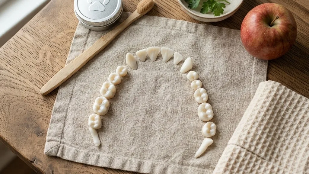
Dog dental chart searches usually start with one blunt question: how many teeth does my dog actually have, and what is going on in there? An adult dog carries permanent teeth, split between the top and the bottom. A puppy runs a smaller set of 28. This guide maps every tooth by type, walks through the modified Triadan numbering that veterinarians write on the chart during an exam, and grades gum disease from PD0 to PD4 using American Animal Hospital Association staging.
If you have ever squinted at a vet report full of numbers, the code cracks open here. A dental map provides the exact location of every issue in your pet’s mouth. For food questions that ride alongside oral health, our breakdown of Showtime Dog Food and its label numbers pairs well with a solid daily routine.
Quick answer: A dog dental chart shows the layout, count, and numbering of a dog’s teeth. An adult dog has 42 permanent teeth (12 incisors, 4 canines, 16 premolars, 10 molars); a puppy has 28 deciduous teeth with no molars. Veterinarians label each tooth using the modified Triadan system, where four quadrants (100, 200, 300, 400) are combined with a two-digit position number. The chart also records gum health, staged PD0 through PD4, so problems get caught early.
How Many Teeth a Dog Has and What Each Type Does
An adult dog has 42 permanent teeth: 12 incisors, 4 canines, 16 premolars, and 10 molars, as documented by the Merck Veterinary Manual. Those 42 split into 20 maxillary teeth on the upper jaw and 22 mandibular teeth on the lower jaw. A puppy carries only 28 deciduous teeth, following the formula two times (I 3/3, C 1/1, P 3/3), which means a puppy has no molars at all.
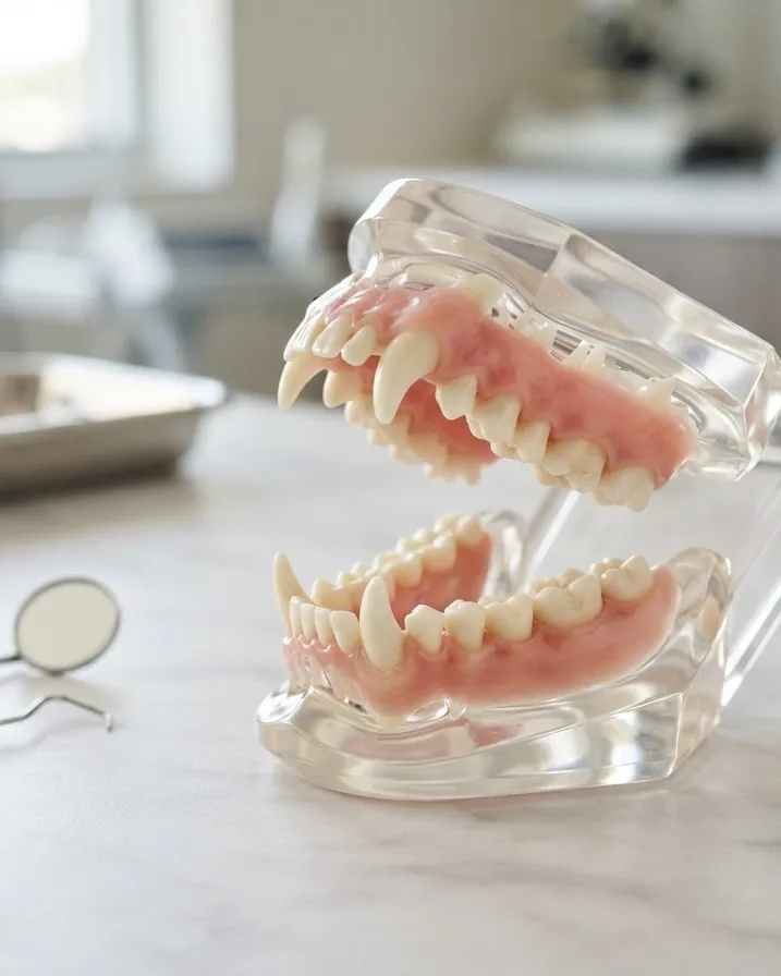
Each of the four tooth types earns its place in the mouth. Reading a dog dental chart starts with recognizing which type sits where, because the numbering system that follows depends entirely on knowing a canine from a premolar. The shape and size of each tooth dictates how many roots anchor it into the jawbone.
The Incisors (12 Teeth)
Incisors form the small front row of teeth between the canines. A dog has six top incisors and six bottom incisors. These teeth have a single root. Dogs use their incisors for delicate work like scraping meat from a bone, grooming their coat to remove burrs or fleas, and picking at small pieces of food. When a dog nibbles gently on your hand, they are using their incisors.
The Canines (4 Teeth)
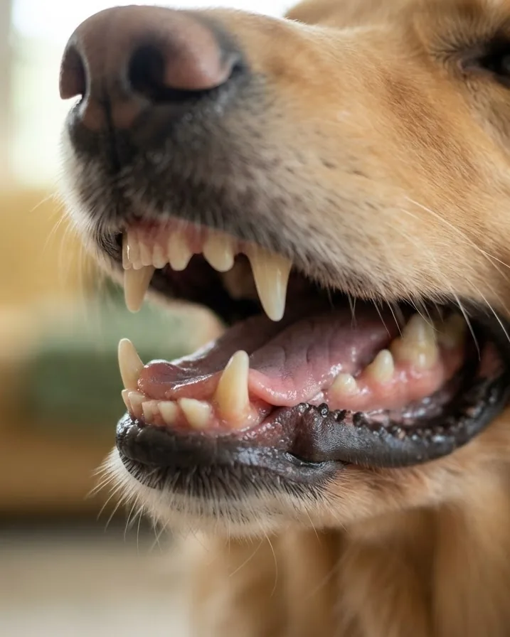
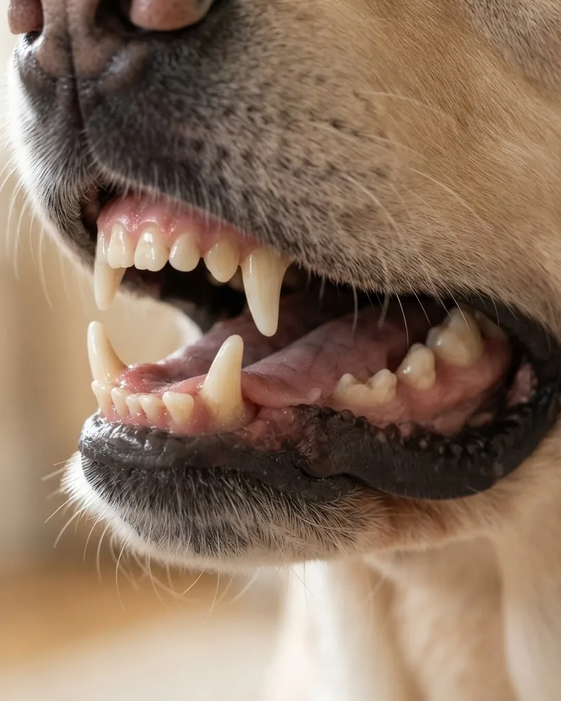
The canines are the four long, curved fangs located just behind the incisors. There are two on the top jaw and two on the bottom. Built for grasping and puncturing, they lock onto objects and hold them tight. Canine teeth have extremely long, thick single roots that extend deep into the nasal cavity area on the upper jaw. Breaking a canine tooth is a major dental event that requires immediate veterinary attention.
The Premolars (16 Teeth)
Premolars are the workhorses just behind the canines. An adult dog has eight on the top and eight on the bottom. They feature sharp, serrated edges that cut like scissors. Dogs tilt their heads to the side to use their premolars to shear through tough material. The upper fourth premolar is massive and takes the brunt of heavy chewing forces.
The Molars (10 Teeth)
Molars are the back grinders. Unlike the other tooth types, the distribution is uneven: adult dogs have four up top (two on each side) and six on the bottom (three on each side). Molars have flat surfaces designed for crushing dry food and hard treats. The uneven top-to-bottom count is the reason the lower jaw carry two extra teeth compared with the upper jaw.
The Modified Triadan System: Reading the Numbers
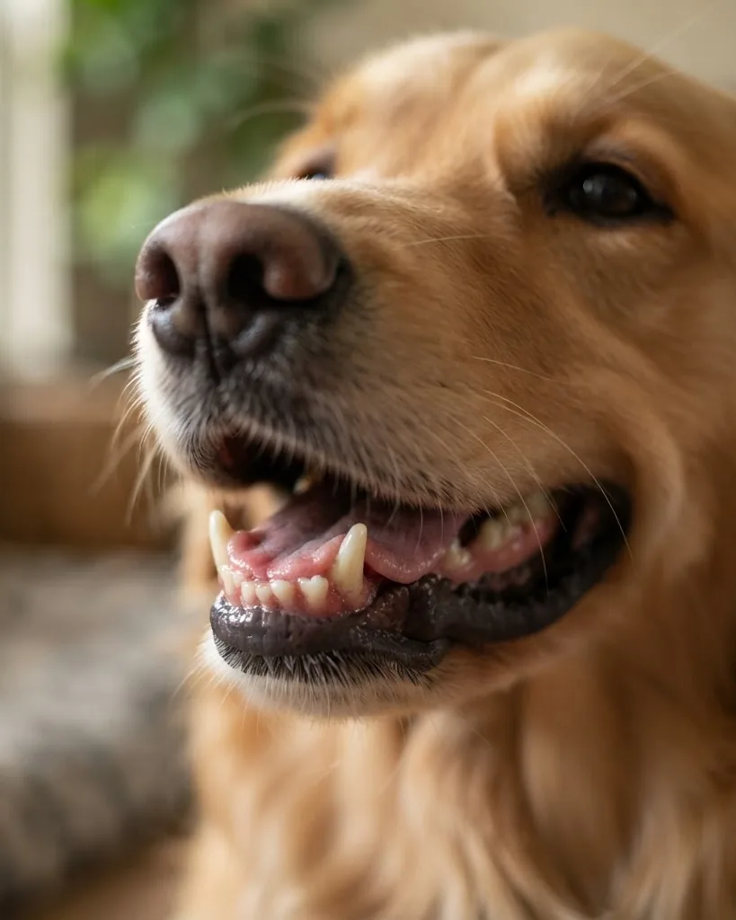
The modified Triadan system is the numbering language every veterinary dental chart speaks, and it turns 42 confusing teeth into a tidy code. The mouth divides into four quadrants. The upper right is the 100 series. The upper left is the 200 series. The lower left is the 300 series. The lower right is the 400 series. Deciduous puppy teeth use the 500 through 800 series instead.
Each tooth then gets a two-digit position number counting back from the midline of the face. The American Veterinary Dental College, which sets this nomenclature, uses two anchor rules that make the whole system readable. First, the canine tooth is always designated as 04. Second, the first molar is always designated as 09.
Missing teeth are simply skipped, never renumbered. If a dog is born without a first premolar, the chart jumps directly to the next expected tooth. The system was introduced by Floyd in the Journal of Veterinary Dentistry in 1991 and remains the worldwide standard. You can confirm the exact naming rules through the Merck Veterinary Manual dentition and nomenclature page.
Complete Adult Dog Dental Chart Numbers
To fully understand your vet’s notes, you need to know exactly which tooth matches which number. Here is the complete breakdown of all 42 permanent teeth across the four quadrants. This section provides a direct translation key for your pet’s medical records.
Upper Right Quadrant (100 Series)
- 101: Upper right first incisor.
- 102: Upper right second incisor.
- 103: Upper right third incisor.
- 104: Upper right canine.
- 105: Upper right first premolar.
- 106: Upper right second premolar.
- 107: Upper right third premolar.
- 108: Upper right fourth premolar (the massive three-rooted carnassial tooth).
- 109: Upper right first molar.
- 110: Upper right second molar.
Upper Left Quadrant (200 Series)
- 201: Upper left first incisor.
- 202: Upper left second incisor.
- 203: Upper left third incisor.
- 204: Upper left canine.
- 205: Upper left first premolar.
- 206: Upper left second premolar.
- 207: Upper left third premolar.
- 208: Upper left fourth premolar (the matching carnassial tooth).
- 209: Upper left first molar.
- 210: Upper left second molar.
Lower Left Quadrant (300 Series)
- 301: Lower left first incisor.
- 302: Lower left second incisor.
- 303: Lower left third incisor.
- 304: Lower left canine.
- 305: Lower left first premolar.
- 306: Lower left second premolar.
- 307: Lower left third premolar.
- 308: Lower left fourth premolar.
- 309: Lower left first molar (the lower carnassial tooth, two roots).
- 310: Lower left second molar.
- 311: Lower left third molar (a very small, single-rooted peg tooth).
Lower Right Quadrant (400 Series)
- 401: Lower right first incisor.
- 402: Lower right second incisor.
- 403: Lower right third incisor.
- 404: Lower right canine.
- 405: Lower right first premolar.
- 406: Lower right second premolar.
- 407: Lower right third premolar.
- 408: Lower right fourth premolar.
- 409: Lower right first molar.
- 410: Lower right second molar.
- 411: Lower right third molar.
Puppy Teeth Versus Adult Teeth: The Eruption Timeline
Puppies and adults run on two completely different dental charts, and the switch happens very fast. A puppy has 28 deciduous teeth. Because they lack molars, their modified Triadan numbers stop early in each quadrant. The puppy quadrants are 500 (upper right), 600 (upper left), 700 (lower left), and 800 (lower right).
Those baby teeth begin erupting at roughly 3 weeks of age. The incisors usually appear first, followed quickly by the needle-like canines. The deciduous premolars erupt afterward. Eventually, a puppy has a fully functional set of sharp baby teeth, ready for weaning onto solid foods.
The permanent set then replaces them in a rapid wave. This replacement process generally begins with the incisors. The adult canines follow. The entire permanent dentition of 42 teeth is usually fully erupted and in place by 6 to 7 months of age.
Retained Deciduous Teeth
One common issue charted by veterinarians is a retained deciduous tooth. This happens when a baby tooth refuses to fall out while the adult version erupts right beside it. This creates a trap for food and plaque. For example, a vet might note a retained 504 right next to a healthy 104. Small breeds like Yorkshire Terriers and Chihuahuas face this problem frequently, and the baby tooth usually requires surgical extraction.
Tooth Roots: Why the Chart Marks Single Versus Multi-Root
A dog dental chart does not just count teeth; it maps how many roots anchor each one, and that detail drives every extraction decision. Knowing root anatomy is necessary for safe surgical planning. The Merck Veterinary Manual details exact root counts for every tooth type. Simple teeth like incisors and canines have a single root. The heavy chewing teeth have more, branching into the jawbone for stability.
The maxillary fourth premolar, numbered 108 and 208, carries three distinct roots. The maxillary first and second molars (109, 110, 209, 210) are also three-rooted. In the lower jaw, the major chewing teeth like the first molar (309, 409) have two massive roots. That anatomy is exactly why pulling a back tooth is a surgical procedure, not a quick tug. Each root must be separated with a high-speed drill and elevated individually, otherwise the jawbone could fracture.
- Single root: All incisors (01-03), all canines (04), first premolars (05), and the bottom third molar (11).
- Two roots: Maxillary second and third premolars (06, 07). Mandibular second, third, and fourth premolars (06, 07, 08). Mandibular first and second molars (09, 10).
- Three roots: Maxillary fourth premolar (08) and maxillary first and second molars (09, 10).
Periodontal Disease: Staging Your Dog’s Gums From PD0 to PD4
Periodontal disease is the primary reason a dog dental chart exists. The disease moves through a predictable sequence: soft plaque hardens into mineralized tartar, the tartar inflames the gum tissue into gingivitis, and untreated gingivitis becomes deep periodontitis. This destroys the bone and ligament attachment holding the teeth in place. The American Veterinary Dental College and the American Animal Hospital Association grade this progression on a strict five-point scale.
Stage 0: Normal (PD0)
PD0 represents a clinically healthy mouth. The gums are firm, pink, and feature a sharp margin where they meet the tooth. There is no bleeding when probed, no plaque accumulation, and no attachment loss. This is the goal of daily home care, though it is rare to see a perfect PD0 in adult dogs without regular professional cleanings.
Stage 1: Gingivitis (PD1)
PD1 marks the onset of gingivitis. The margin of the gum becomes red and swollen, and plaque covers the teeth. However, there is zero attachment loss. The bone and ligaments remain perfectly intact. This is the only stage of periodontal disease that is 100 percent reversible with a professional cleaning and strict home care.
Stage 2: Early Periodontitis (PD2)
PD2 indicates less than 25 percent attachment loss. The gums bleed easily when probed, and the vet will note shallow pockets forming around the roots. Tartar is usually thick. Bad breath becomes noticeable. While the lost bone cannot be replaced, professional treatment can halt the disease here and save the tooth.
Stage 3: Moderate Periodontitis (PD3)
PD3 sits at 25 to 50 percent attachment loss. The gums pull away from the teeth, exposing the sensitive roots. The teeth may begin to wobble slightly. Probing reveals deep pockets filled with bacteria. At this stage, advanced surgical techniques or extractions are necessary. A dog in PD3 is likely experiencing chronic pain while eating.
Stage 4: Advanced Periodontitis (PD4)
PD4 is severe, with more than 50 percent of the attachment structure destroyed. The teeth are visibly loose and roots are fully exposed. Chronic infection is present, posing a risk to the dog’s internal organs as bacteria enter the bloodstream. Teeth at this stage almost always require immediate extraction.
Probing Depths and Dog Breed Sizes
Staging is not guesswork based on how bad the teeth look. A veterinarian measures the sulcus, the small groove between tooth and gum, using a graduated periodontal probe. Per World Small Animal Veterinary Association guidance, a normal canine sulcus reads less than 3 mm deep. Anything deeper signals an abnormal pocket where the attachment has pulled away from the root.
Probing depth is read against body size, since a large dog’s mouth tolerates deeper pockets before the same percentage of root loss applies. Using AVDC criteria, size brackets generally split as large (over 25 kg), medium (10 to 25 kg), and small (under 10 kg). A 5 mm pocket on a Great Dane might only indicate PD2, while a 5 mm pocket on a Chihuahua indicates catastrophic PD4 bone loss.
Warning Signs That Send You to the Vet
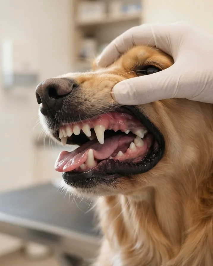
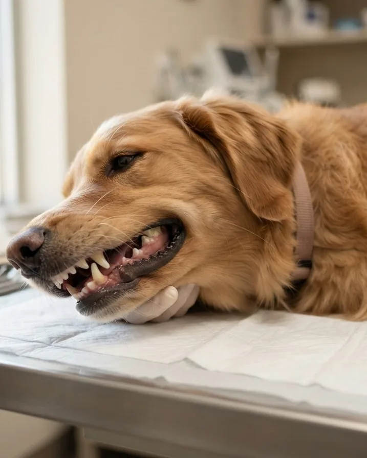
Before relying on any home routine, you must know the clinical red flags. Persistent bad breath is not normal dog physiology; it is a sign of rotting tissue or bacteria. Red or bleeding gums, a cracked tooth, a wobbly tooth, and any tooth that has gone missing without explanation all warrant a professional exam.
None of these issues should be managed by home brushing alone. Brushing a fractured tooth causes severe pain. The American Society for the Prevention of Cruelty to Animals warns that untreated dental disease can lead to jaw fractures from bone destruction and organ damage from chronic systemic infection, which is why early flags matter so much.
What Vets Record During a Dental Charting Exam
A veterinary charting exam fills in far more than simple tooth counts. Understanding what gets recorded helps you read the discharge sheet. During a full assessment under anesthesia, the veterinary team probes and notes sulcus depth at six different points around every single tooth. They check for tooth mobility, enamel fractures, resorptive lesions, and gum recession.
They also check for furcation exposure. The furcation is the area where multiple roots join the crown. If bone loss exposes this junction, the chart marks it as a severe staging factor. The American Animal Hospital Association’s 2019 Dental Care Guidelines for Dogs and Cats lay out a strict 12-step protocol for every dental procedure.
The AAHA 12-Step Protocol
The AAHA guidelines demand a comprehensive approach to oral health. The steps include a preliminary awake exam, induction of general anesthesia, subgingival scaling (cleaning below the gumline), supragingival scaling (cleaning above the gumline), polishing to smooth the enamel, subgingival irrigation, and full-mouth intraoral radiographs.
The remaining steps involve detailed periodontal probing and charting, formulating a diagnosis, planning the treatment, performing necessary surgery or extractions, and providing the owner with home care instructions. Dental radiographs accompany the physical probing because the vast majority of periodontal disease hides below the gumline in the jawbone.
Why Anesthesia-Free Scaling Fails the Chart
The American Veterinary Dental College holds a firm position against anesthesia-free dental scaling, a cosmetic procedure offered by some groomers. The college argues it cannot clean below the gumline where the disease actually lives. Scraping the visible crown makes the tooth look white, but it leaves the dangerous subgingival plaque fully intact.
Furthermore, a dog must be intubated and anesthetized to safely take dental X-rays and probe the 3 mm sulcus depths without pain. You can read the college’s reasoning in its position statement on dental scaling without anesthesia, and the full medical protocol in the 2019 AAHA Dental Care Guidelines resource center.
Home Care: Using Your Chart to Improve Health
Home dental care is the single biggest lever an owner controls, and it hinges on consistency more than intensity. Daily brushing is the ideal target; brushing 2 to 3 times per week is a reasonable minimum that still slows plaque before it mineralizes into hard tartar. The one non-negotiable rule is toothpaste selection.
Use a canine formula only, never human toothpaste. Human pastes contain fluoride and often xylitol, an artificial sweetener that is highly toxic to dogs. Beyond the brush, the Veterinary Oral Health Council awards a seal of approval to dental products that meet strict plaque- and tartar-control standards. Checking for the VOHC seal filters empty marketing claims from scientifically tested products.
Skin and coat problems sometimes travel alongside oral neglect during grooming lapses. While you build a daily brushing habit, our guides to choosing a hypoallergenic dog shampoo and spotting a dog hot spot before it spreads round out a full at-home care routine.
Dog Versus Cat: A Quick Dental Comparison
Dogs and cats do not share a dental chart, and the gap is bigger than most owners assume. An adult dog has 42 permanent teeth; an adult cat has just 30. That 12-tooth difference reflects diet and jaw design, since cats are obligate carnivores with absolutely no need for the flat grinding molars that dogs carry.
The probing thresholds differ drastically too. A healthy canine sulcus reads under 3 mm, while a healthy feline sulcus sits strictly under 1 mm. This means the exact same 2 mm pocket is perfectly normal in a Golden Retriever but represents dangerous bone loss in a Siamese cat. The numbers, the counts, and the healthy baselines do not cross over.
| Feature | Adult Dog | Adult Cat |
|---|---|---|
| Permanent teeth | 42 | 30 |
| Maxillary (top) teeth | 20 | 16 |
| Mandibular (bottom) teeth | 22 | 14 |
| Healthy sulcus depth | Under 3 mm | Under 1 mm |
| Puppy/kitten teeth | 28 deciduous | 26 deciduous |
Frequently Asked Questions
How many teeth does an adult dog have?
An adult dog has 42 permanent teeth, made up of 12 incisors, 4 canines, 16 premolars, and 10 molars, according to the Merck Veterinary Manual. They divide into 20 teeth on the upper jaw and 22 on the lower jaw. Puppies have a smaller set of 28 deciduous teeth with no molars at all.
What does the number 108 mean on a dental chart?
Under the modified Triadan system used by the American Veterinary Dental College, 108 is the upper right fourth premolar. The first digit, 1, marks the upper right quadrant, and 08 is the tooth’s position counting back from the midline. It is a massive three-rooted shearing tooth, which is why fractures there are common and extractions are heavily involved.
What does a missing number on the chart mean?
If your vet’s chart skips from 304 to 306, it means the 305 tooth is missing. The Triadan system assigns a permanent number to every possible position. If a tooth never erupted, fell out, or was previously extracted, the number is skipped to maintain the standard naming convention for the rest of the mouth.
How deep should my dog’s gum pockets be?
A healthy sulcus, the groove between tooth and gum, measures less than 3 mm deep in dogs, per World Small Animal Veterinary Association guidance. Anything deeper suggests a periodontal pocket where the attachment has pulled away. Your veterinarian measures this with a probe during an exam and stages it from PD0 to PD4.
Is anesthesia-free dental cleaning safe for dogs?
The American Veterinary Dental College advises against anesthesia-free dental scaling because it cannot clean below the gumline, where periodontal disease actually develops. It may polish the visible crown while leaving the real problem untreated. A proper cleaning under anesthesia allows probing, radiographs, and subgingival scaling that a conscious procedure simply cannot achieve safely.
How often should my dog get a dental exam?
The 2019 AAHA Dental Care Guidelines recommend an oral exam every 6 months for a healthy dog, moving to every 3 to 6 months if periodontitis is already present. The American Veterinary Medical Association also recommends a comprehensive veterinary oral check at least once a year as a strict minimum for every dog.
When do puppies get their adult teeth?
Puppy deciduous teeth begin erupting around 3 weeks of age, and the full adult set of 42 teeth is usually in place by 6 to 7 months. During the changeover you may find shed baby teeth or see red, swollen gums. If a puppy is still missing expected adult teeth, have a veterinarian check for retained roots.
Why does my dog have double fangs?
Double fangs occur when a deciduous baby canine fails to fall out while the permanent adult canine erupts beside it. This condition is known as a retained deciduous tooth. The baby tooth (usually numbered 504 or 604) traps debris against the adult tooth (104 or 204) and usually requires surgical removal by a vet.
Can a dog eat dry food with missing teeth?
Yes, dogs adapt remarkably well to missing teeth. Even dogs that have had full-mouth extractions due to severe stage 4 periodontal disease can learn to eat dry kibble by swallowing the small pieces whole, just as they would in the wild. Eliminating the source of chronic dental pain is always better than keeping infected teeth for chewing.
Putting the Chart to Work
A dog dental chart is only useful if it changes what you do at the sink and at the clinic. Learn the counts of 42 adult teeth and 28 puppy teeth. Recognize the four tooth types, and decode the Triadan numbers so a medical report stops being a foreign language. Keep track of which teeth have multiple roots, as these are the hardest to extract if they become infected.
Then act on the health layer: brush most days, watch for bad breath and bleeding gums, and keep the yearly exam that the American Veterinary Medical Association recommends. If digestive or other severe symptoms show up alongside oral problems, our guide to a dog throwing up white foam and its red flags can help you decide when a symptom is an emergency. None of this replaces your veterinarian, who remains the only one qualified to stage disease and decide on surgical treatment.